
“fMRI Visualization of Multiple Functional Areas” by Hardenbergh
Conference:
Type(s):
Entry Number: 108
Title:
- fMRI Visualization of Multiple Functional Areas
Presenter(s)/Author(s):
Abstract:
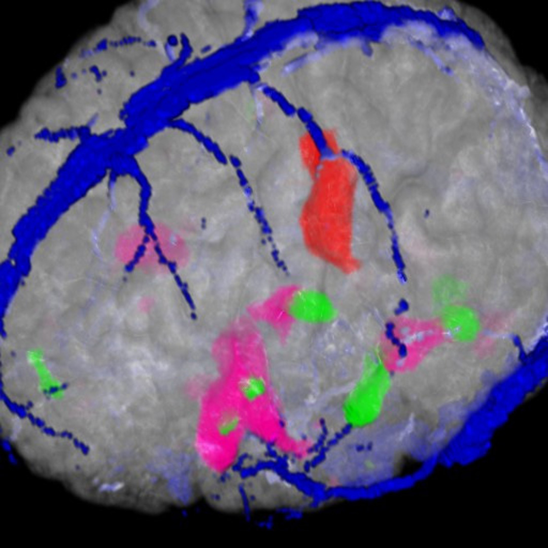
Functional Magnetic Resonance Imaging (fMRI) displays regions of the brain activated for mental functions. Some of the identified functional areas are: language generation and acquisition, motor control, touch, visual processing, and many more. The ability to plan around these areas is very useful in neurosurgical planning.
fMRI takes advantage of the change in blood flow to active regions of the brain. With increased flow, there is a local reduction in oxygen unloaded deoxyhemoglobin, which is paramagnetic. These regions of increase can be identified in the strong magnetic field used for MR imaging.
The reconstructed scan data consists of a volume dataset for each activation area as well as the anatomy and vasculature (blood vessels). We display the anatomy and vasculature to provide context for the activations. In hardware that is two texture reads and two dependent (LUT) reads. Naively, each activation volume requires a texture lookup and two dependent reads. Display of three activation areas requires 13 texture reads, 3 serial pixel subtractions and 4 additions. This is not possible on the VP1000 [Wu 2003]. Also, memory requirements, complexity and performance on current graphics processors leads us to look for alternative solutions to displaying multiple activation areas.
References:
1. Wu, Y., Bhatia, V., Lauer, H., Seiler, L. 2003. Shear-image ray casting volume rendering. In Proceedings of the 2003 symposium on Interactive 3D graphics, ACM Press.
Acknowledgements:
Image rendered the TeraRecon VolumePro 1000. Data courtesy of MGH 3D Imaging Service. Application knowledge: Shirley Miller.