
“Virtual Dog Head: Using 3D Models to Teach Complex Veterinary Anatomy” by Viehdorfer, Nemanic, Mills and Bailey
Conference:
- SIGGRAPH 2014
-
More from SIGGRAPH 2014:


Type(s):
Title:
- Virtual Dog Head: Using 3D Models to Teach Complex Veterinary Anatomy
Presenter(s):
Description:
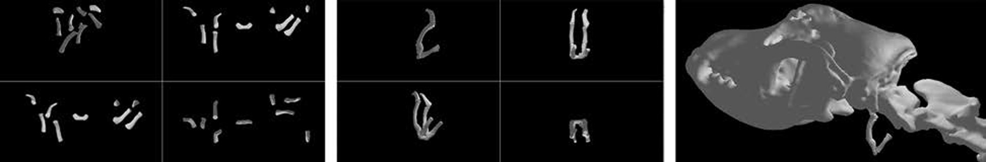
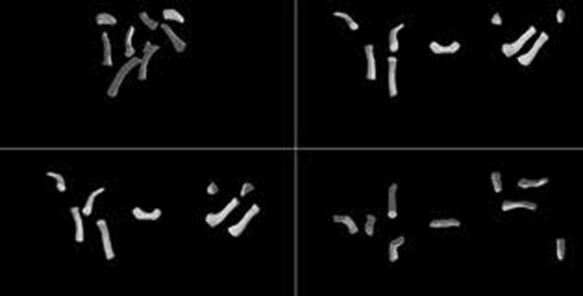
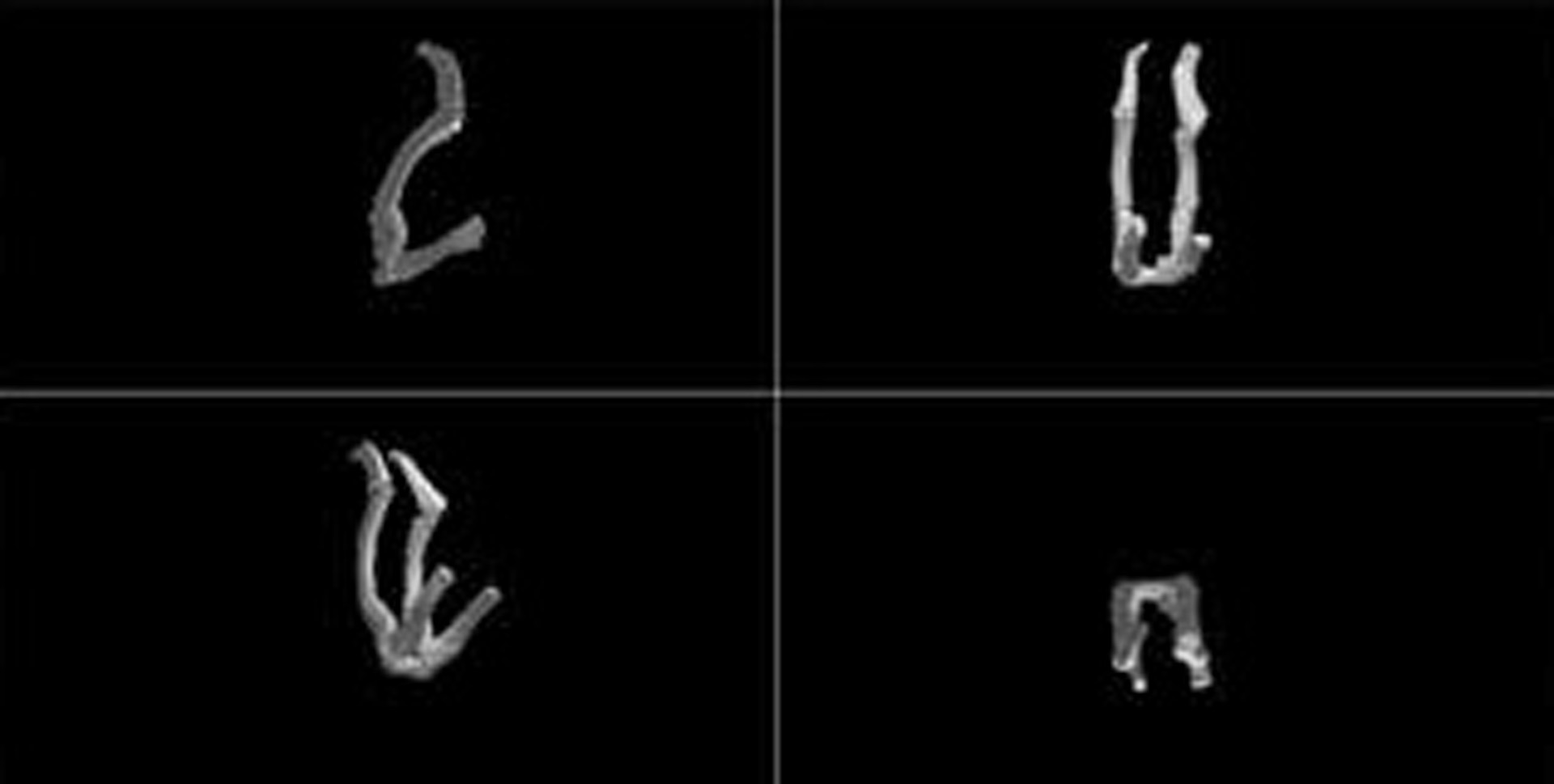
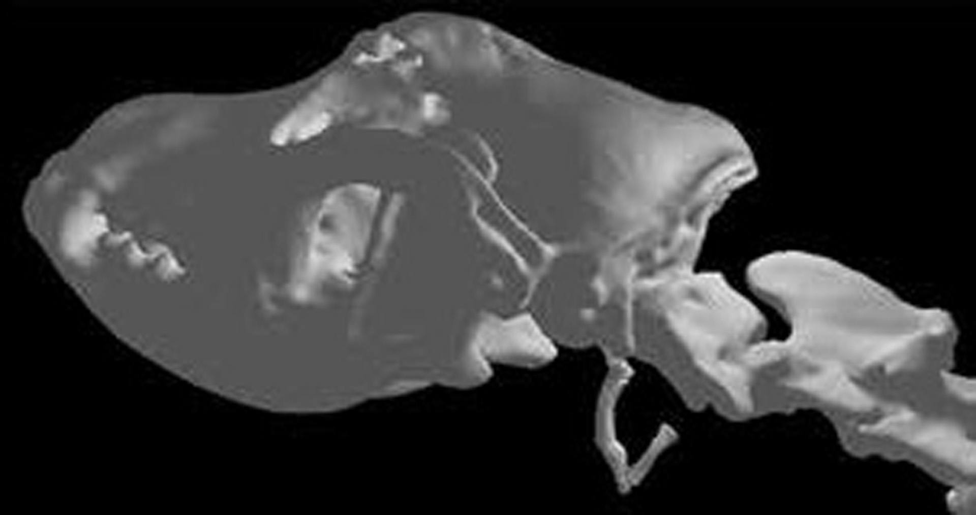
Oregon State University veterinary medicine faculty recently observed that fourth year veterinary medical students consistently struggled during their clinical rotations in cases that required accurate and detailed recall of the complex anatomical relationships in the head region of a dog. These structures were difficult to see and understand since they were hidden behind muscle and tissue and often destroyed or damaged during dissection.
This work presents a new method of interactive learning through the incorporation of 3D computed tomography (CT) scan-generated models of the hyoid and larynx apparatus in dogs with OpenGL and the Oculus Rift. It will also be used as a review resource for fourth year veterinary medicine students on clinical rotations.