John McGhee: Integrity: structure and surface
Artist(s):
Title:
- Integrity: structure and surface
Exhibition:
- SIGGRAPH 2007: Global Eyes
-
More artworks from SIGGRAPH 2007:


Creation Year:
- 2007
Size:
- 30 inches x 40 inches x 1 inch
Category:
Artist Statement:
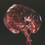
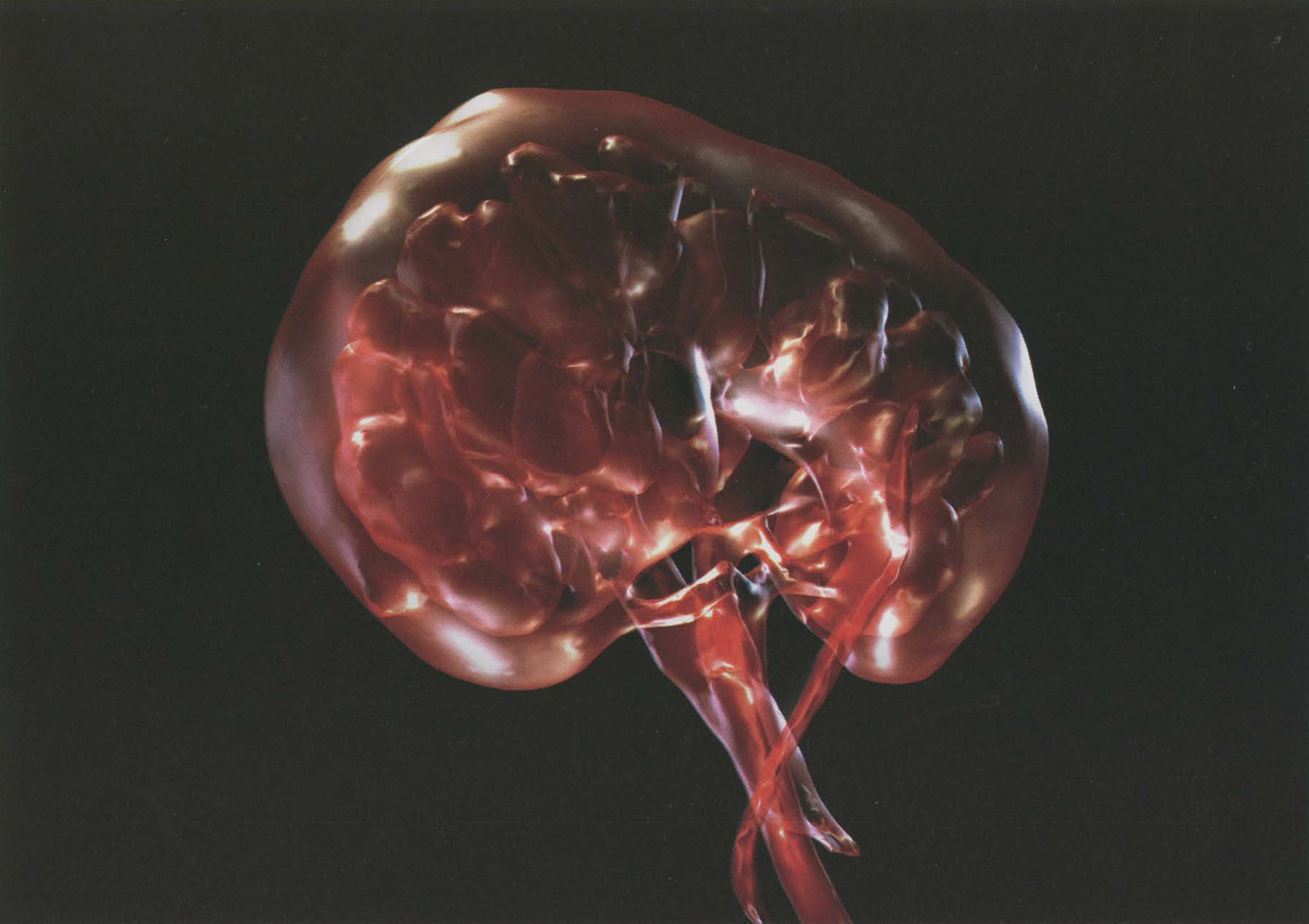
These images explore my own interest in beauty, structure, and harmony within the human vascular system, in particular the flow of blood through the human kidney. Medical technologies such as magnetic resonance imaging (MRI) allow clinicians to see ever deeper into our body spaces for the purpose of diagnosis. However, interpretation is restricted to the eye of the trained specialist in a clinical or scientific environment. In this work, 3D computer visualisation techniques were used to create a hybrid image that bridges scientific medical data and artistic imaginative vision to create a new aesthetic. These images were created as part of an ongoing collaboration with the Department of Clinical Radiology at Ninewells Hospital, Dundee, Scotland. My artwork has focused on the visualisation of the human vascular system and methods of emotion-based interpretation of MRI scan data. The reasoning behind these particular images was to expose and communicate the fragility of the human kidney. The intention was to suggest an internal jewel-like quality to anatomy, highlighting an inner space otherwise hidden from the human eye. My goal was to produce an image with a degree of sensibility in an attempt to bring the patient closer to the nature and beauty of inner human structure, and also as a means of penetrating the complexity of arterial disease.
Technical Information:
The starting point for the creation of these images is MRI data. In the first instance, DICOM-image-slice data were exported from a Siemens Avanto MRI scanner located in
Ninewells Hospital. These cross-sectional slices are acquired during an angiogram procedure performed on a patient being scanned for a vascular condition known as renal artery stenosis. This diagnostic scan involves a contrast agent being injected into the vascular system during the scan procedure. From the DICOM files, the data are imported into a medical 3D reconstruction package (Materialise Mimics) and, through a process of thresholding and segmentation, an ISO surface is extracted. After it is cleaned up, the 3D geometry
is imported into AutoDesk Maya 7, and the anatomy is creatively interpreted with the addition of shaders, lighting, and rendering.