
“Computer graphics to illustrate the development of a human embryo for professional medical education” by Kakusho, Minekura, Minoh, Mizuta, Nakatsu, et al. …
Conference:
Type(s):
Interest Area:
- Application
Title:
- Computer graphics to illustrate the development of a human embryo for professional medical education
Session/Category Title: Visualizing Humans
Presenter(s)/Author(s):
Abstract:
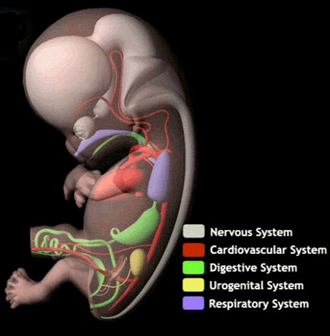
This sketch describes three dimensional (3D) computer graphics (CG) produced to illustrate the development of a human embryo for education in embryology, which is one of the basic subjects in professional medical education (Fig. 1). Although similar CG have already been produced for TV programs, they are insufficient in precision for professional medical education.
References:
1. B. R. Smith, D. S. Huff, and G. A. Johnson. 1999. Magnetic resonance imaging of embryos: An internet resource for the study of embryonic development. Computerized Medical Imaging and Graphics 23, 33–40.
2. R. O’Rahilly, and F. Muller. 1987. Developmental Stages in Human Embryos. Carnegie Institution of Washington.
ACM Digital Library Publication:
- Computer graphics to illustrate the development of a human embryo for professional medical education