
“Ultrasound Visualization with the Sonic Flashlight” by Shelton, Stetten and Chang

Conference:
Experience Type(s):
E-Tech Type(s):
Title:
- Ultrasound Visualization with the Sonic Flashlight
Entry Number: 82
Organizer(s)/Presenter(s):
Description:
From the discovery of X-rays over a century ago, clinicians have been presented with a wide assortment of imaging modalities yielding maps of localized structure and function within the patient. Some imaging modalities are tomographic, meaning that the data are localized into voxels, rather than projected along lines of sight as with conventional X-ray images. Tomographic modalities include magnetic resonance (MR), computerized tomography (CT), ultrasound, and others. Tomographic images, with their spatially distinct voxels, are essential to our present work.
New techniques to display tomographic images have lagged behind the development of imaging modalities themselves. In the practice of medicine, the standard method of viewing an image is still to examine a photographic film or an electronic screen rather than to look directly into the patient. The ability to view images at their actual locations within the patient could have a broad impact on the diagnosis and treatment of disease by providing in situ guidance for invasive procedures.
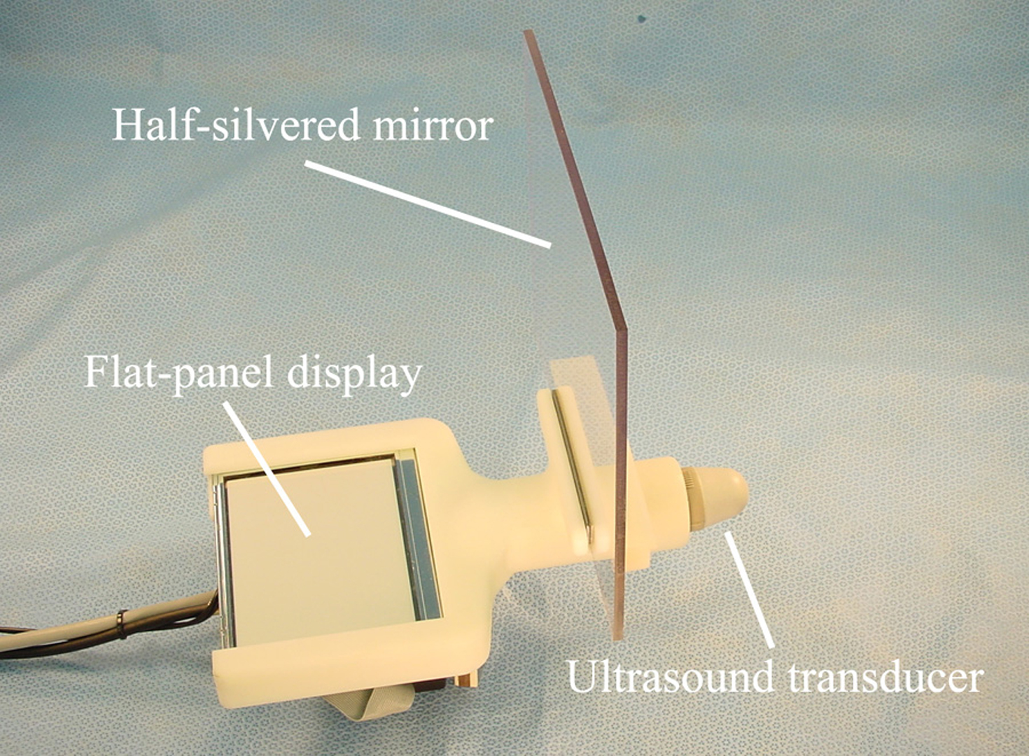
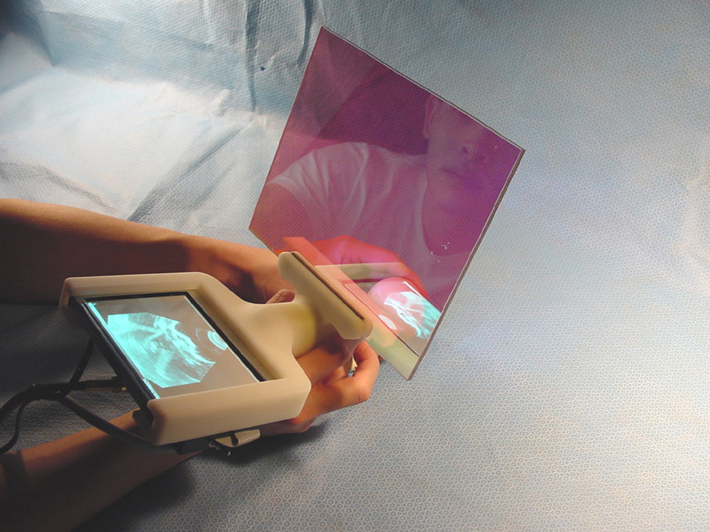
We have developed a new method of guidance that displays an image in situ within the patient in real time. The method, which we call real time tomographic reflection (RTTR), employs a half-silvered mirror to combine a direct view of the patient with a reflection from the display of an imaging device. The reflected image appears to be floating within the patient at its actual location, and the user perceives this 3D effect using natural, viewpoint independent, stereoscopic vision. Unlike previous attempts at such a visual merger, RTTR does not require tracking the patient or the viewer and involves no head-mounted apparatus. By presenting the patient, the operator’s hands, the invasive tool, and the image in a single perceptual environment, the operator may take full advantage of natural hand-eye cooperation.
Our present implementation of RTTR uses ultrasound as its imaging modality and is named the Sonic Flashlight because the reflected ultrasound image appears to emanate from the transducer. Because of its small size, it can be used single-handed, leaving the other hand free to perform medical procedures such as ultrasound guided needle biopsy. Unlike many other tomographic visualization techniques, such as those involving head tracking, third-party observers not directly manipulating the device also view the ultrasound in its correct physical location, and the unit may be passed between multiple personnel during a procedure without requiring recalibration or transfer of bulky external equipment.
Preliminary tests of the Sonic Flashlight have demonstrated the ability to hit targets using natural hand-eye coordination inside common ultrasound phantoms such as water-filled balloons and turkey breasts. Recently, we have collaborated with an ophthalmologist to test the ability of the Sonic Flashlight to guide a needle during a procedure known as retrobulbar injection, during which a long needle is used to inject a therapeutic substance behind the eyeball. Further clinical testing is planned for a wide variety of procedures, including catheter insertion and tumor biopsy.