
“Flattened anatomy for interactive segmentation & measurement” by Gering, Potter and Foo
Conference:
Type(s):
Title:
- Flattened anatomy for interactive segmentation & measurement
Presenter(s)/Author(s):
Abstract:
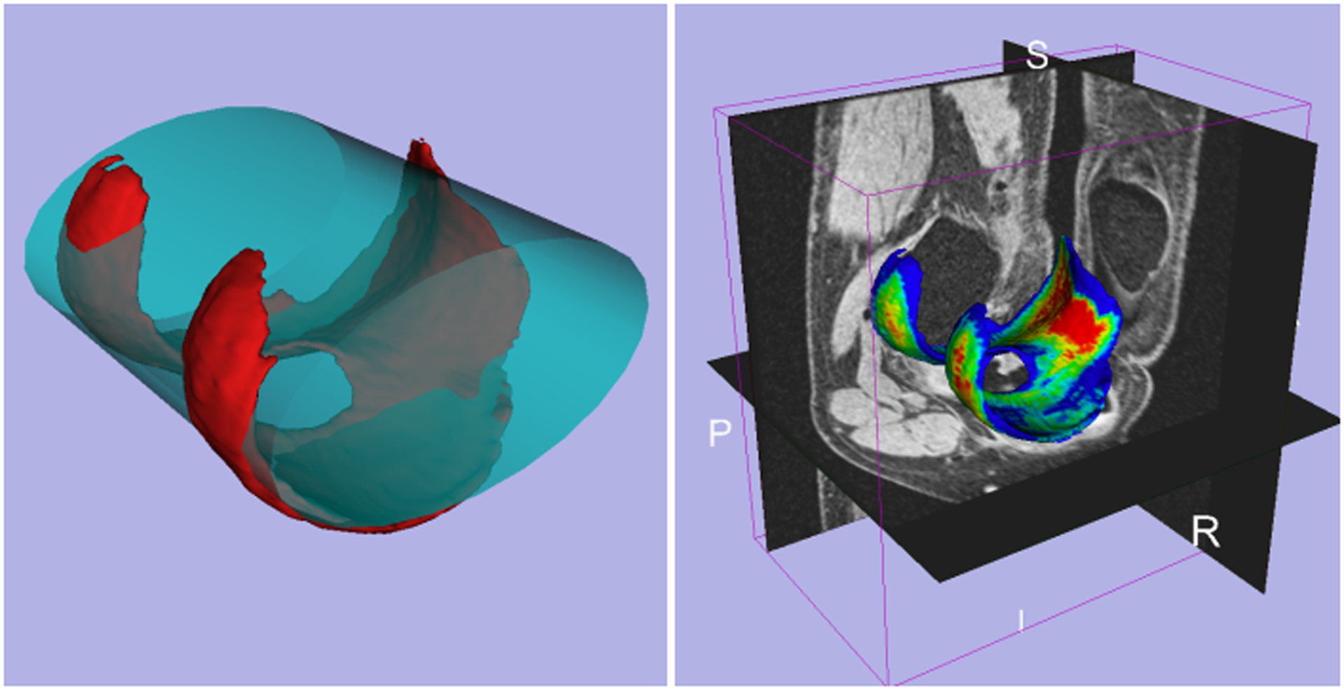
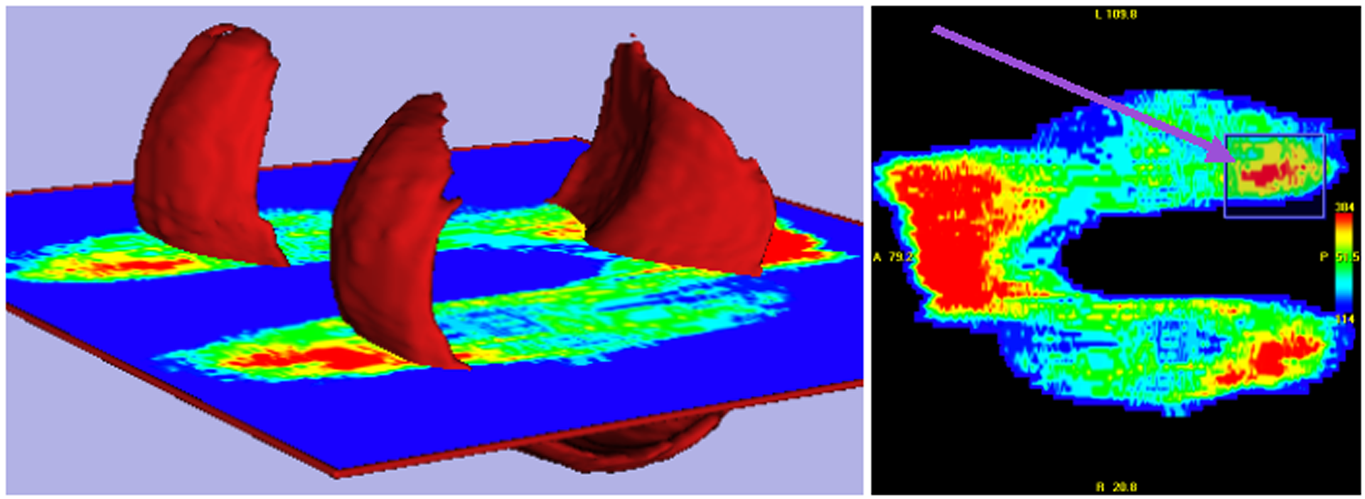
Anatomical surfaces can be extracted from a volume of medical imagery through segmentation, which is the process of labeling image voxels according to the tissue type represented. Many anatomical surfaces can be described as 2-D manifolds embedded within 3-D space. The manifolds could be linear, such as a plane, or non-linear, such as a curved sheet. In some applications, it would be desirable for the user to be able to interact with the manifold by drawing upon it in some way. The purpose of this drawing could be to perform quantitative measurements such as to measure distances, surface areas, or volumes. The purpose could also be to analyze local properties of the 3-D surface at specific locations, where these properties include thickness and curvature.
References:
1. Bartroli, A. V., Wegenkittl, R., Konig, A., Groller, E., Sorantin, E. Virtual Colon Flattening. VisSym 2001, p. 127–136.
2. Halir, R., Flussr, J. Numerically Stable Direct Least Squares Fitting Of Ellipses. Proc. 6th WCSG, 1998.