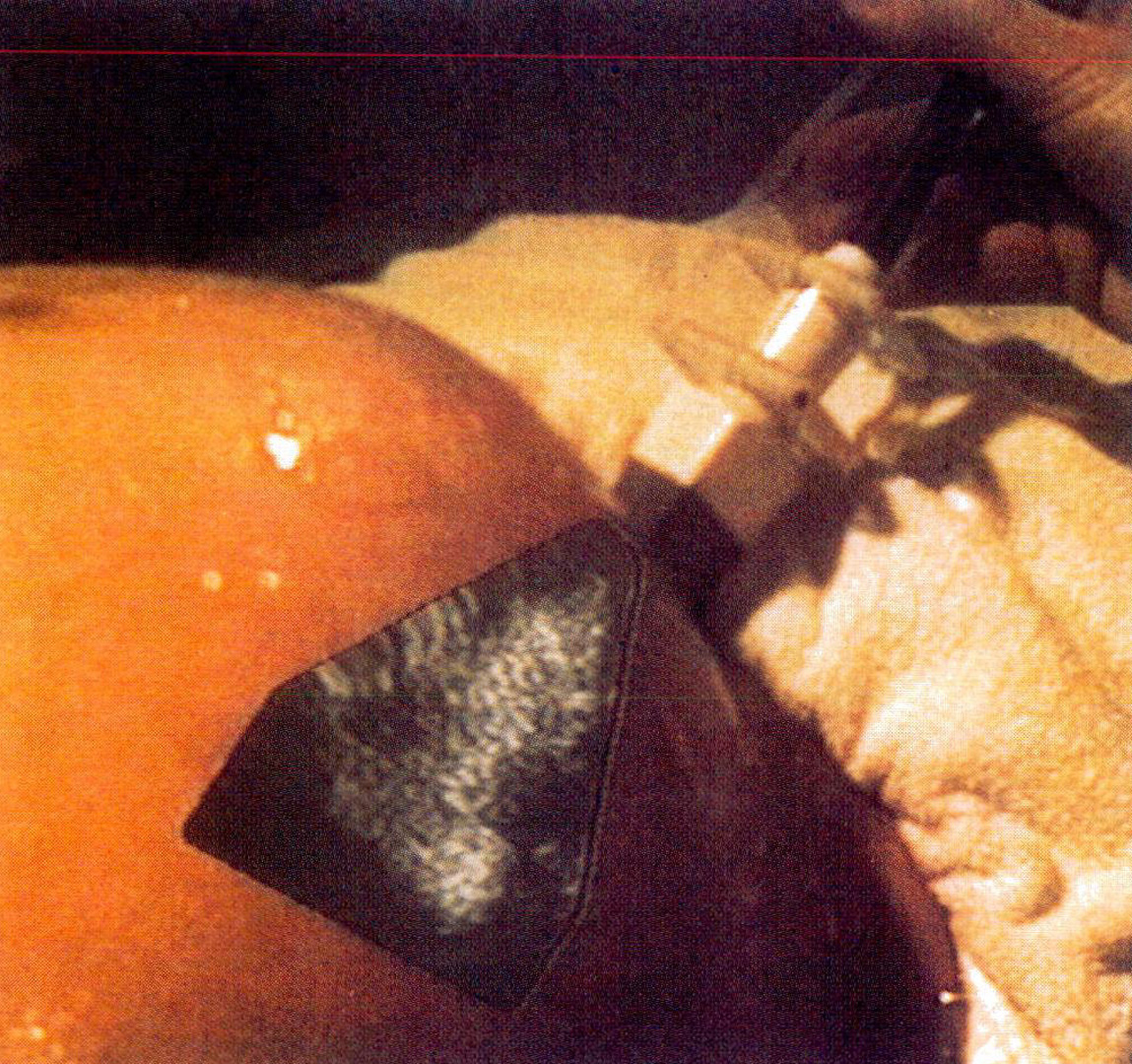
“Merging virtual objects with the real world: seeing ultrasound imagery within the patient” by Bajura, Fuchs and Ohbuchi
Conference:
Type(s):
Title:
- Merging virtual objects with the real world: seeing ultrasound imagery within the patient
Presenter(s)/Author(s):
Abstract:
No abstract available.
References:
1. Brinkley, J. F., Moritz, W.E., and Baker, D.W. “Ultrasonic Three-Dimensional Imaging and Volume From a Series of Arbitrary Sector Scans.” Ultrasound in Med. &Biol., 4, pp317-327.
2. Collet Billon, A., Philips Paris Research Lab. Personal Communication.
3. Fornage, B. D., Sneige, N., Faroux, M.J., and Andry, E. “Sonographic appearance and ultrasound guided fine-nee~Jle aspiration biopsy of brest carcinomas smaller than 1 cm . lournal of Ultrasound in Medicine, 9, pp559-568.
4. Fuchs, H., GoldFeather, j., Hultiquist, J.P., Spach, S., Austin, J., Brooks, Jr., F.P. Eyles, J., and Poulton, J. “Fast Spheres, Textures, Transparencies, and Image Enhancements in Pixel Planes.” Computer Graph&’s (Proceedings of SIGGRAPH’ 85 ), 19(3), ppl 11-120.
5. Fuchs, H., Poulton, J., Eyles, J., Greer, T., Goldfeather, j., Ellsworth, D., Molnar. S., and israel, L. “‘Pixel Planes 5: A Heterogeneous Multiprocessor Graphics System Using Processor-Enhanced Memories.” Computer Graphit’s (Proceedings of SIGGRAPH’ 89), 23(3), pp79-88.
6. Galyean, T. A., and Hughes, J.F. “‘Sculpting: An Interactive Volumetric Modeling Technique.” ComputerGraphits (Prot’eedings of SIGGRAPH’ 89), 25(4 ), pp267-274.
7. Geiser, E. A. Ariet, M., Conetta, D.A., Lupkiewicz, S.M., Christie, L.G., and Conti, C.R. “Dynamic three-dimensional echocardiographic reconstruction of the intact human left ventricle: Technique and initial observations in patients.”Amerit’an Heart journal, 10316), pp1056-1065.
8. Geiser, E. A., Christie, L.G., Conetta, D.A., Conti, C.R., and Gossman, G.S. “Mechanical Arm for Spatial Registration of Two-Dimensional Echographic Sections.” Cathet. Cariovas~’. Diagn., 8, pp89- I01.
9. Ghosh, A., Nanda, C.N., and Maurer, G. “‘Three- Dimensional Reconstruction of Echo-Cardiographics Images Using The Rotation Method.” Ultrasound in Med. & Biol.,S(6), pp655-661.
10. Haddad, R. A., and Parsons, T.W. Digital Signal Processing, Theory, Appli~’ations, and Hardware. New York, Computer Science Press.
11. Harris, R. A., Follett, D.H., Halliwell, M, and Wells, P.N.T. “Ultimate limits in ultrasonic imaging resolution.” Ultrasound in Medicine and Biology, 17(6), pp547-558.
12. Hildreth, E. C. “The Detection of Intensity Changes by Computer and Biological Vision Systems.” Computer Vision, Graphites, and Image Processing, 22, ppl-27.
13. Hottier, F., Philips Paris Research Lab. Personal Communication.
14. Hottier, F., Collet Billon, A. 3D Echography: Status and Perspective. 3D Imaging in Medicine. Springer-Verlag. pp2 i-41.
15. Itoh, M., and Yokoi, H. “‘A computer-aided threedimensional display system for ultrasonic diagnosis of a breast tumor.” Ultrasonics,, pp261-268.
16. King, D. L., King Jr., D.L., and Shao, M.Y. “Three- Dimensional Spatial Registration and interactive Display of Position and Orientation of Real-Time Ultrasound Images.” Journal of Ultrasound Med, 9, pp525-532.
17. Lalouche, R. C., Bickmore, D., Tessler, F., Mankovich, H.K., and Kangaraloo, H. “Three-dimensional reconstruction of ultrasound images “SPIE’89, Medical imaging, pp59-66.
18. Leipnik, R. “The extended entropy uncertainty principle.” Info. Control, 3, ppl 8-25.
19. Levoy, M. “Display of Surface from Volume Data.” IEEE CG&A, 8(5), pp29-37.
20. Levoy, M. “A Hybrid Ray Tracer for Rendering Polygon and Volume Data.” IEEE CG&A, 10(2), pp33-40.
21. Liang, J., Shaw, C., and Green, M. “On Temporal- Spatial Realism in the Virtual Reality Environment.” User Interface Software and Technology, 1991, Hilton Head, SC., U.S.A., pp19-25.
22. Lin, W., Pizer, S.M., and Johnson, V.E. “Surface Estimation in Ultrasound Images.” Information Processing in Medical Imaging 1991, Wye, U.K., Springer-Verlag, Heidelberg, pp285- 299.
23. Matsumoto, M., lnoue, M., Tamura, S., Tanaka, K., and Abe, H. “Three-Dimensional Echocardiography for Spatial Visualization and Volume Calculation of Cardiac Structures.” J. Clin. Ultrasound, 9, pp157-165.
24. McCann, H. A., Sharp, J.S., Kinter, T.M., McEwan, C.N., Bariliot, C., and Greenleaf, J.F. “Multidimensional Ultrasonic Imaging for Cardiology.” Proc.IEEE, 76(9), pp1063- 1073.
25. Mills, P. H., and Fuchs, H. “3D Ultrasound Display Using Optical Tracking.” First Conference on Visualization for Biomedical Computing, Atlanta, GA, IEEE, pp490-497.
26. Miyazawa, T. “A high-speed integrated rendering for interpreting multiple variable 3D data.” SPIE, 1459(5),
27. Molnar, S., Eyles, J., and Poulton, J. “PixelFlow: High-Speed Rendering Using Image Composition.” Computer Graphics (Proceedings of SIGGRAPH’92), ((In this issue)),
28. Moritz, W. E., Pearlman, A.S., McCabe, D.H., Medema, D.K., Ainsworth, M.E., and Boles, M.S. “An Ultrasonic Techinique for Imaging the Ventricle in Three Dimensions and Calculating Its Volume.” IEEE Trans. Biota. Eng., BME-30(8), pp482-492.
29. Nakamura, S. “Three-Dimensional Digital Display of Ultrasonograms.” IEEE CG&A, 4(5), pp36-45.
30. Ohbuchi, R., and Fuchs, H. “Incremental 3D Ultrasound Imaging from a 2D Scanner.” First Conference on Visualization in Biomedical Computing, Atlanta, GA, IEEE, pp360- 367.
31. Ohbuchi, R., and Fuchs, H. “Incremental Volume Rendering Algorithm for Interactive 3D Ultrasound Imaging.” information Processing in Medical Imaging 1991 (Lecture Notes in Computer Science, Springer-Verlag), Wye, UK, Springer- Verlag, pp486-500.
32. Pini, R., Monnini, E., Masotti, L., Novins, K. L., Greenberg, D. P., Greppi, B., Cerofolini, M., and Devereux, R. B. “Echocardiographic Three-Dimensional Visualization of the Heart.” 3D Imaging in Medicine, Travemiinde, Germany, F 60, Springer-Verlag, pp263-274.
33. Polhemus. 3Space Isotrak User’s Manual.
34. Raichelen, J. S., Trivedi, S.S., Herman, G.T., Sutton, M.G., and Reichek, N. “Dynamic Three Dimensional Reconstruction of the Left Ventricle From Two-Dimensional Echocardiograms.” Journal. Amer. Coll. of Cardiology, 8(2), pp364-370.
35. Robinett, W., and Rolland, J.P. “A Computational Model for the Stereoscopic Optics of a Head-Mounted Display.” Presence, 1(1), pp45-62.
36. Sabeila, P. “A Rendering Algorithm for Visualizing 3D Scalar Fields.” Computer Graphics (Proceedings of SIGGRAPH’88), 22(4), pp51-58.
37. Shattuck, D. P., Weishenker, M.D., Smith, S.W., and yon Ramm, O.T. “Explososcan: A Parallel Processing Technique for High Speed Ultrasound Imaging with Linear Phased Arrays.” JASA, 75(4), pp1273-1282.
38. Smith, S. W., Pavy, Jr., S.G., and yon Ramm, O.T. “High-Speed Ultrasound Volumetric Imaging System – Part I: Transducer Design and Beam Steering.” IEEE Transaction on Ultrasonics, Ferroelectrics, and Frequency Control, 38(2), ppl00-108.
39. Stickels, K. R., and Warm, L.S. “An Analysis of Three-Dimensional Reconstructive Echocardiography.” Ultrasound in Med. & Biol., 10(5), pp575-580.
40. Thijssen, J. M., and Oosterveld, B.J. “Texture in Tissue Echograms, Speckle or Information ?” Journal of Ultrasound in Medicine, 9, pp215-229.
41. Tomographic Technologies, G. Echo-CT.
42. Upson, C., and Keeler, M. “VBUFFER: Visible Volume Rendering.” ACM Computer Graphics (Proceedings of SIGGRAPH’ 88 ), 22(4), pp59-64.
43. von Ramm, O. T., Smith, S.W., and Pavy, Jr., H.G. “High-Speed Ultrasound Volumetric Imaging System – Part II: Parallel Processing and Image Display.” IEEE Transaction on Ultrasonics, Ferroelectrics, and Frequency Control, 38(2), ppl09-115.
44. Ward, M., Azuma, R., Bennett, R., Gottschalk, S., and Fuchs, H. “A Demonstrated Optical Tracker with Scalable Work Area for Head-Mounted Display Systems.” 1992 Symposium on Interactive 3D Graphics, Cambridge, MA., ACM, pp43-52.
45. Wells, P. N. T. Biomedical ultrasonics. London, Academic Press.


