
“Lensless Stereo Microscopic Imaging” by Zimmerman and Smith
Conference:
Experience Type(s):
Title:
- Lensless Stereo Microscopic Imaging
Entry Number: 16
Organizer(s)/Presenter(s):
Description:
Introduction
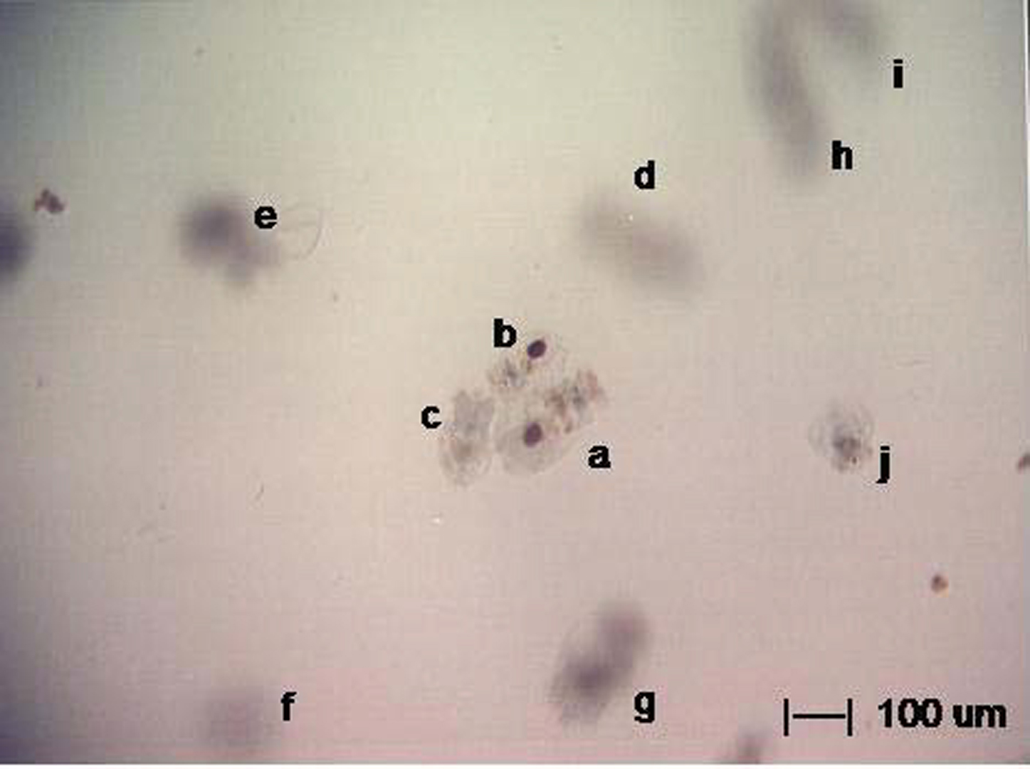
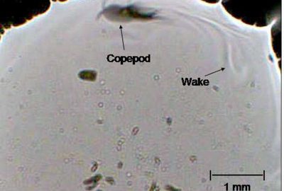
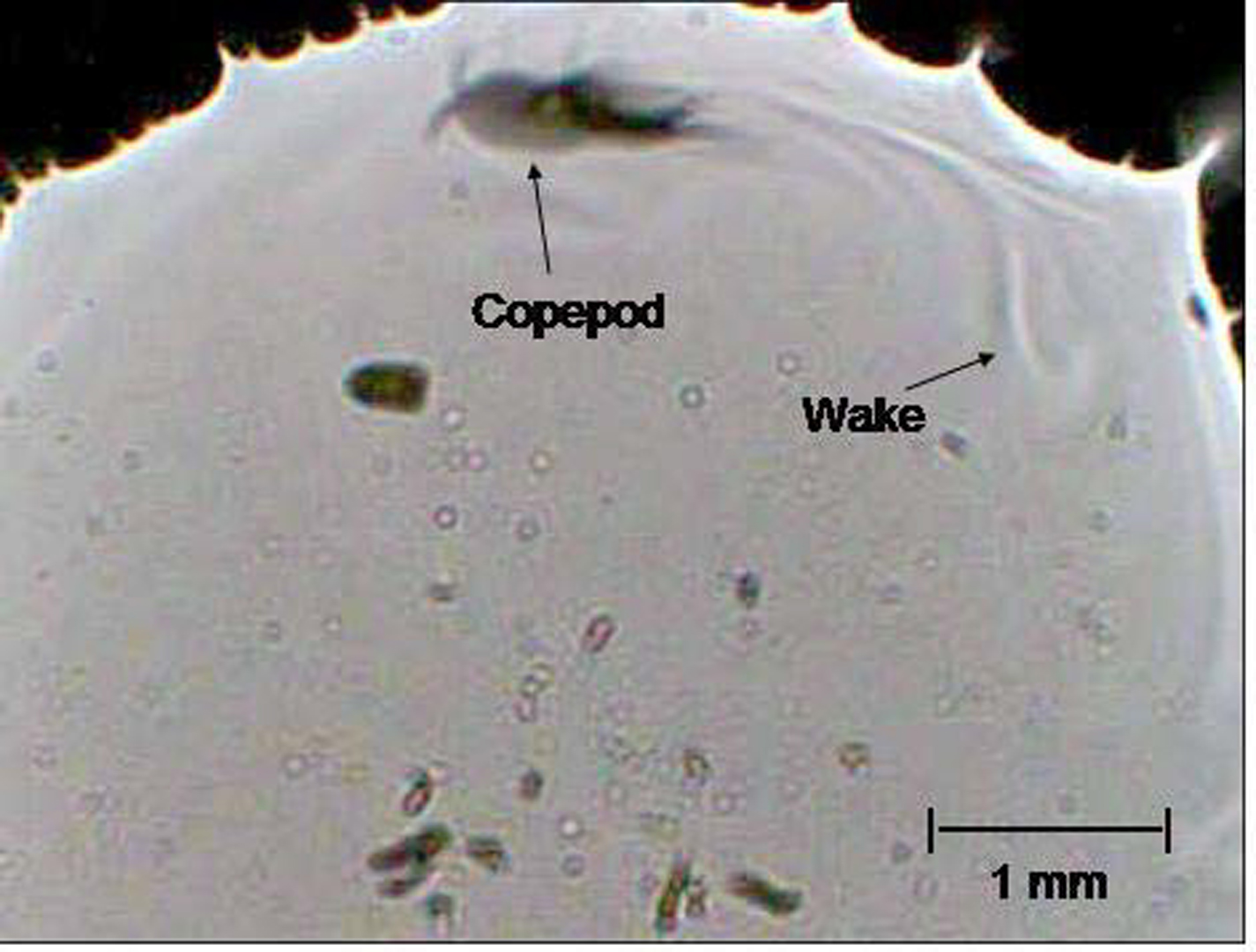
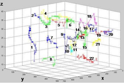
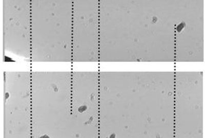
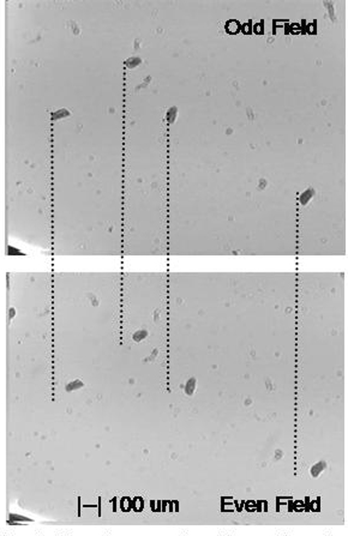
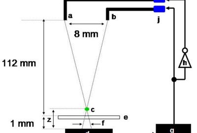
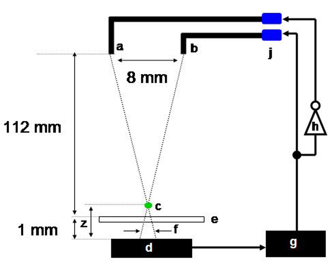
A simple inexpensive high-contrast stereo microscopic is constructed from a single video imager sensor. Two field- synchronous LEDs illuminate the subject creating disparity. The stereo microscope outputs standard field-sequential 3D video and is compatible with commercial head mounted displays and LCD shutter glasses.
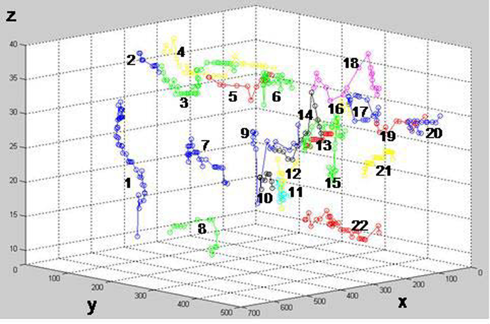
The microscope has no lens, no focus, a large depth of field and no distortion between the stereo channels, reducing depth calculation to a single one-dimensional sliding correlation per object.
The easy-to-use microscope provides students and teachers with a fast and convenient way to share images of microorganisms. The microscope is compact and robust, well suited for remote sensing. The focus-free low-cost design makes it practical for a computer to continuously monitor and manage an array of microscopes for high-throughput biological experiments and research.
Other Information:
References
BERG, H. C., BROWN, D.A. 1972 Chemotaxis in Escherichia coli analyzed by Three-dimensional Tracking. Nature, 239:5374, 500-504.
CHAROY, C. 1995. Modification of the swimming behaviour of Brachionus calyciflorus (Pallas) according to food environment and individual nutritive state. Journal Hydrobiologia, 313-314:1, 197-204.
COULON, P.Y., CHARRAS, J.P., CHASSE, L., CLEMENT, P, CORNILLAC, A., LUCIANI, A., WURDAK, E., 1983. An experimental system for the automatic tracking and analysis of rotifer swimming behaviour. Journal Hydrobiologia 104:1, 197-202.
GARCIA-SUCERQUIA, J., XU, W., JERICHO, S. K., KLAGES, P., JERICHO, M.H., KREUZER, H.J., 2006. Digital in-line holograhic microscopy. Applied Optics 45, 836-850.
HENG, H., ERICKSON, D., PSALTIS, D., YANG C., 2005.A new imaging method: optofluidic microscopy. Invited Talk, SPIE Optics East, Boston MA.
HOELING, B., FERNANDEZ, A., HASKELL,R., HUANG, E., MYERS, W., PETERSON, D., UNGERSMA, S., WANG, R. , WILLIAMS, M., FRASER, S., 2000. An optical coherence microscope for 3-dimensional imaging in developmental biology, Optics Express 6, 136-146.
RASKAR, R., TAN, K., FERIS, R., YU, J. TURK, M. 2004. Non-photorealistic camera: Depth edge detection and stylized rendering using multi-flash imaging. ACM Transactions on Graphics. 23, 3, 679-688.
REED MARICULTURE 2007, 520 McGlincy Lane #1, Campbell, CA 95008 www.reed-mariculture.com/rotifer/
YUFERAL, M., PASCUALL, E., OLIVARES, J.M. 2005. Factors Affecting Swimming Speed in the Rotifer Brachionus plicatilis. Journal Hydrobiologia, 546:1, 375-380.
Additional Images:
-
- 2007 ETech Zimmerman: Lensless Stereo Microscopic Imaging
-
- 2007 ETech Zimmerman: Lensless Stereo Microscopic Imaging
-
- 2007 ETech Zimmerman: Lensless Stereo Microscopic Imaging
-
- 2007 ETech Zimmerman: Lensless Stereo Microscopic Imaging
-
- 2007 ETech Zimmerman: Lensless Stereo Microscopic Imaging
Acknowledgements:
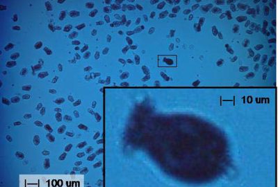
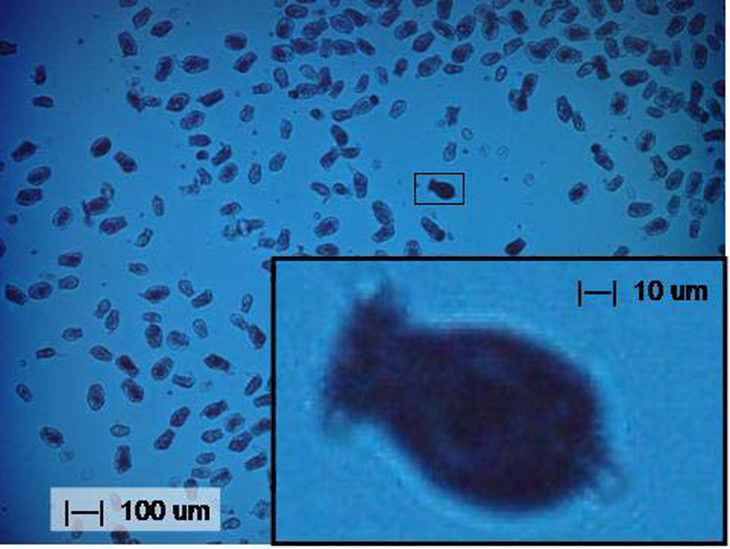
The authors would like to thank Eugene Delenia of IBM Research for providing the optical microscope image of rotifer and Reed Mariculture (Campbell, CA) for providing rotifer and copepods for our research. The lensless stereo microscope is patent pending.