
“3-D computer arts in clinical radiology” by McGhee, Houston and Chesson
Conference:
Type(s):
Title:
- 3-D computer arts in clinical radiology
Presenter(s)/Author(s):
Abstract:
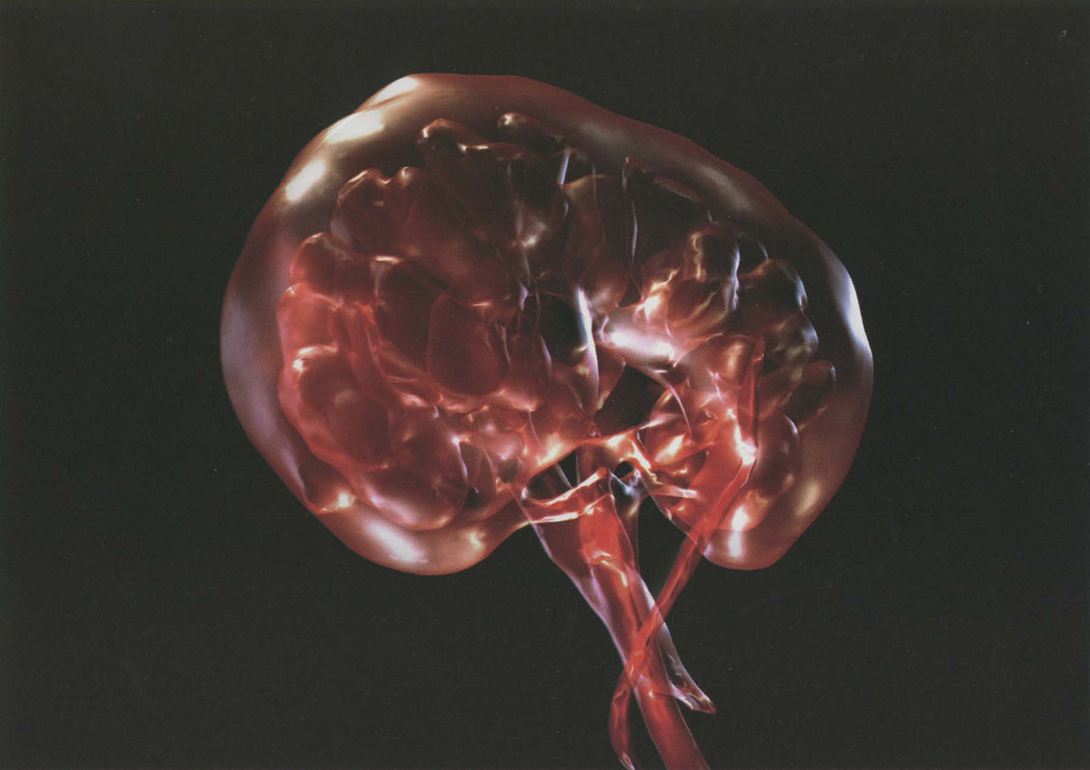
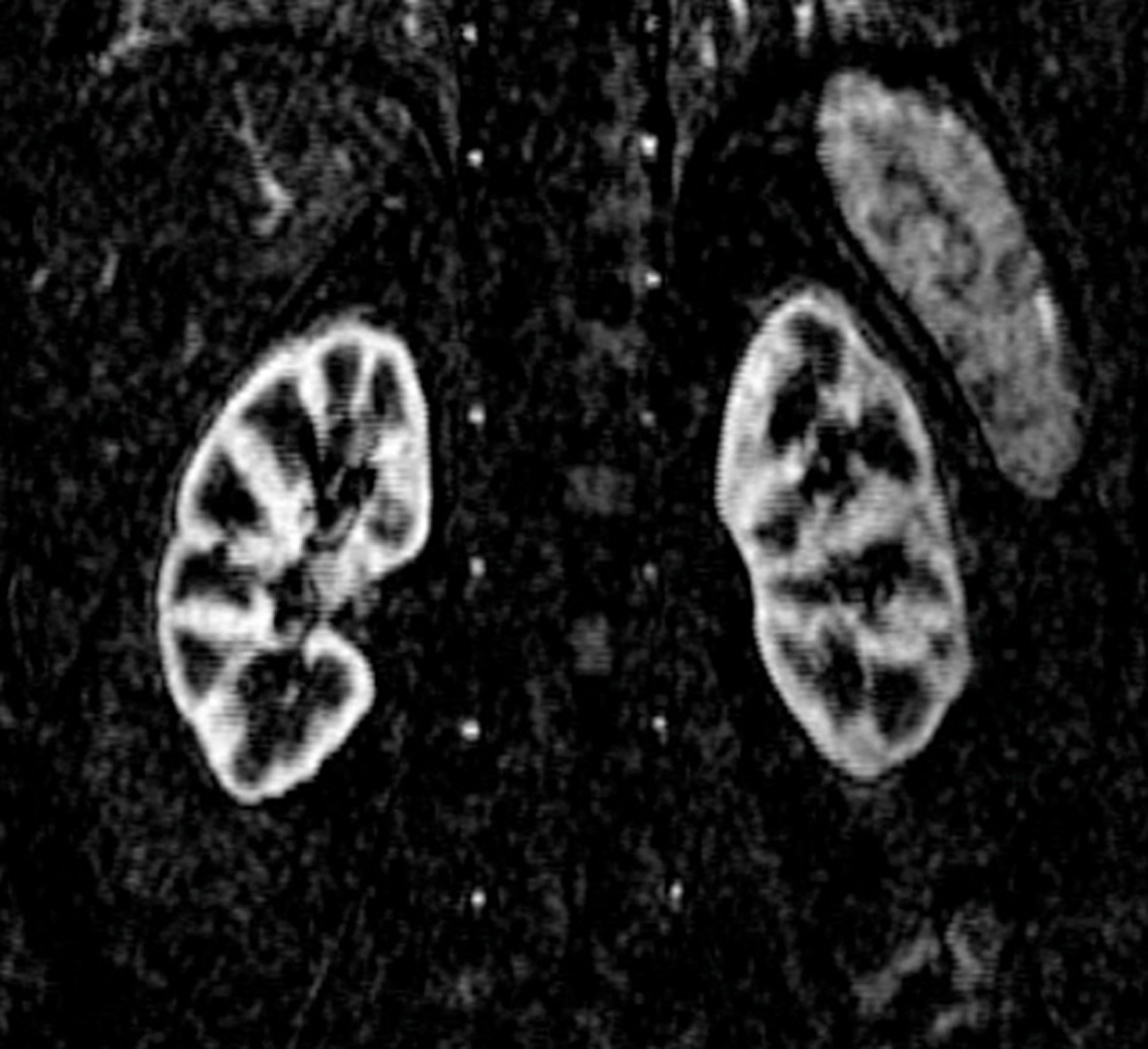
There is increasing evidence to suggest patients want to be better informed about their own disease diagnosis [Chesson et al. 2002; Broadbent et al. 2004]. Based on ongoing collaborative work between the School of Media Arts & Imaging and Clinical Radiology, Ninewells Hospital, Dundee, a need has been identified for 3-D visualisation strategies to improve disease understanding among patient groups. Scientific imagery such as Magnetic Resonance Imaging (MRI) and Computed Tomography (CT) allows clinicians to image ever deeper into our body spaces for the purpose of clinical diagnosis. However, interpretation is restricted to the eye of the trained medic within a clinical or scientific context. Captured as a series of 2-D slices (Figure 1), MRI imagery, while useful to a Radiologist, can cause confusion and misunderstanding when presented to patients and their families.
References:
1. Chesson, R. A., Mckenzie, G. A. & Mathers, S. A. (2002) What Do Patients Know About Ultrasound, CT and MRI? Clinical Radiology, 57, 477.
2. Broadbent, E., Petrie, K., Ellis, C., Ying, J., and Gamble, G. (2004) A picture of health – myocardial infarction patients’ drawings of their hearts and subsequent disability: A longitudinal study. Journal of Psychosomatic Research 2004;57:583–58